- Undergraduate
Bachelor's Degrees
Bachelor of ArtsBachelor of EngineeringDual-Degree ProgramUndergraduate AdmissionsUndergraduate Experience
- Graduate
Graduate Experience
- Research
- Entrepreneurship
- Community
- About
-
Search

Ryan Halter
Professor of Engineering
Program Area Lead: Biomedical Engineering
Adjunct Associate Professor of Surgery, Geisel School of Medicine
Technical Associate Director, Center for Precision Health and Artificial Intelligence (CPHAI)
Professor Halter discusses his research developing medical imaging devices and systems for monitoring, diagnosing, and treating disease.
Research Interests
Biomedical instrumentation; electrical impedance tomography and spectroscopy; medical imaging; tissue bioimpedance; cancer detection technologies; traumatic brain injury; medical robotics
Education
- BSc, Engineering Science and Mechanics, Pennsylvania State University 1999
- MSc, Engineering Mechanics, Pennsylvania State University 2001
- PhD, Biomedical Engineering, Dartmouth 2006
Awards
- Society of Critical Care Medicine's Gold Snapshot Award, 2021
- New Investigator Award in Prostate Cancer Research: DoD CDMRP, 2009–2012
- Research Robotic Fellowship: Intuitive Surgical, Inc, 2009–2011
Professional Activities
- Member, Institue of Electrical & Electronic Engineers (IEEE)
- Member, International Society for Electrical Bio-Impedance (ISEBI)
- Member, Dartmouth's Committee for the Protection of Human Subjects (CPHS)
Startups
RyTek Medical
Founder and CEO
Founder and CEO
SynchroHealth
Co-Founder
Co-Founder
Research Projects
-
Electrical Impedance Imaging: Enabling deep space missions through medical imaging and diagnosis of the long-term physiological effects of space travel
Electrical Impedance Imaging: Enabling deep space missions through medical imaging and diagnosis of the long-term physiological effects of space travel
Goal: to provide an imaging tool to effectively monitor the long-term physiological effects
of deep space and automate diagnosis, enabling crew members to be proactive in the
event of injury. More specifically, we are designing an integrated US-EIT system and
demonstrate proof-of-concept of US-EIT for enhanced ultrasound imaging capabilities of
deep internal bleeding.
-
Electrical impedance tomography in pulmonary and cardiac applications
Electrical impedance tomography in pulmonary and cardiac applications
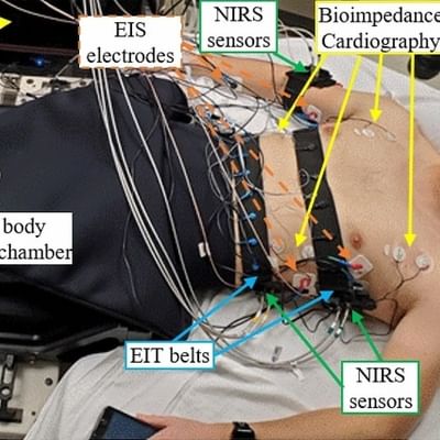
The most common application, so far, of electrical impedance tomography (EIT) has been pulmonary monitoring of patients. In particular, we have been pursuing EIT for cardiac output monitoring (a related application) and EIT as a surrogate for pulmonary function tests.
-
Combined whole breast ultrasound and electrical impedance tomography
Combined whole breast ultrasound and electrical impedance tomography
Ultrasound is a supplemental screening technique that has good sensitivity in dense breasts, is inexpensive, and is widely available, but unfortunately, it has high rates of false positives. Electrical impedance tomography (EIT) is a second attractive modality that is low-cost and has shown promise for cancer detection and in differentiating fibrocystic tissues from other tissues. Combining automated whole breast ultrasound (ABUS), which is a recent improvement to standard ultrasound, with EIT may significantly reduce the number of false positives of ABUS. If successful, the combined ABUS/EIT system could become an important screening technology for women with dense breasts.
-
Enabling technologies for effective image-guided surgical navigation in trans-oral cancer surgery
Enabling technologies for effective image-guided surgical navigation in trans-oral cancer surgery
Throat cancers have been increasing in incidence worldwide. Despite advances in surgical and non-surgical management of these cancers, treatment continues to be associated with significant functional and cosmetic morbidity. More minimally invasive trans-oral surgical (TOS) approaches have reduced treatment morbidity and complications. However, one drawback of TOS is the difficulty in intraoperatively assessing tumor extent and locating major vascular structures. Surgical navigation with image guidance has shown improved safety and efficacy with other surgical procedures; however it is currently not feasible in TOS due to the soft tissue and airway deformation that occurs with placement of instruments needed to access the throat, thus rendering preoperative scans unusable in the intraoperative setting.
The overarching objective of our research is to develop enabling technologies that allow for surgical navigation with image guidance for TOS. Our research strategy is to acquire intraoperative imaging during TOS in order to develop models of upper aerodigestive tract deformation that reflect the intraoperative state. This, in turn, would allow for registration of preoperative images to the intraoperative state. We are well equipped to solve this problem due to the unique intraoperative CT and MR imaging resources available at the Dartmouth Center for Surgical Innovation. We have successfully developed a 3D printable polymer laryngoscopy system which, unlike standard metal laryngoscopes, is CT and MRI compatible. We have also acquired preliminary intraoperative imaging data during laryngoscopy procedures. The next steps in this research will be to further quantify and characterize tissue deformation that occurs during TOS and ultimately develop a surgical navigation platform for trans-oral procedures.
-
Therapy monitoring
Therapy monitoring
Therapy monitoring is an important emerging application of imaging modalities. These and other current research topics include:
- near-infrared imaging of brain tissue;
- near-infrared spectroscopy for diagnosing peripheral vascular disease;
- electrical impedance spectroscopy for radiation therapy monitoring;
- magnetic resonance elastography for detecting brain or prostate lesions; to follow the progression of diabetic damage in the foot; and to answer basic questions of wave propagation in tissue;
- microwave imaging spectroscopy for hyperthermia therapy monitoring, brain imaging, and detection of early-stage osteoporosis;
- electrical impedance tomography for monitoring traumatic brain injury progression and therapy.
-
Combined ultrasound and electrical impedance tomography (EIT)
Combined ultrasound and electrical impedance tomography (EIT)
Combined ultrasound and electrical impedance tomography (EIT) puts 3-D ultrasound imaging together with EIT data in a co-registered volume. EIT relies on the mathematical processing of impedance data collected non-invasively from patients to reconstruct the 3-D distribution of the electrical properties of the tissues inside the patient. Combining ultrasound and EIT has the potential to greatly improve the quality and spatial resolution of the reconstructed electrical properties.
-
Electrical impedance imaging for prostate cancer screening
Electrical impedance imaging for prostate cancer screening
Electrical impedance imaging for prostate cancer screening is the process of imaging non-invasively the electrical properties (conductivity and permittivity) of the prostate and its vicinity using electrodes mounted onto an intracavitary probe.
-
Electrical impedance imaging for breast cancer screening
Electrical impedance imaging for breast cancer screening
Electrical impedance imaging for breast cancer screening is the process of imaging the electrical property (conductivity and permitivity) of tissue using electrodes located on the body surface. This project is one branch of the larger effort to develop innovative technologies for breast cancer detection.
-
Electrical bioimpedance
Electrical bioimpedance
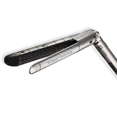
Electrical bioimpedance measurements of tissue provide significant levels of contrast between benign and malignant pathologies due to the vastly different morphologies occurring between tissue types. Focal sensing or mapping of these properties can provide clinicians useful information regarding the extent and severity of diseases like cancer. Our group is currently developing technologies to couple bioimpedance sensors to clinical devices including: 1) intraoperative instruments for use in assessing surgical margins during tumor resection; and 2) standard biopsy needles for use in providing real-time pathological assessment of tissue.
Selected Publications
- Halter RJ, Schned AR, Heaney JA, Hartov A, Paulsen KD, "Electrical properties of prostatic tissues: I. single frequency admittivity properties," Journal of Urology, 182:1600-1607, 2009.
- Halter RJ, Schned AR, Heaney JA, Hartov A, Paulsen KD, "Electrical properties of prostatic tissues: II. Spectral admittivity properties," Journal of Urology, 182:1608-1613, 2009.
- Halter RJ, Zhou T, Meaney PM, Hartov A, Barth, Jr RJ, Rosenkranz KM, Wells WA, Kogel CA, Borsic A, Rizzo EJ, Paulsen KD "Correlation of in vivo and ex vivo tissue dielectric properties to validate electromagnetic breast imaging: initial clinical experience," Physiological Measurement, 30:S121-S136, 2009.
- Halter RJ, Hartov A, Paulsen KD, "Video Rate Electrical Impedance Tomography of Vascular Changes: Preclinical Development," Physiological Measurement, 29(3):349-364, 2008.
- Halter RJ, Hartov A, Paulsen KD, "A broadband high frequency electrical impedance tomography system for breast imaging," IEEE Transactions on Biomedical Engineering, 55(2):650-659, 2008.
Patents
- System and method for forming a cavity in soft tissue and bone | 12256948
- Remote-sensing, bluetooth-enabled resistance exercise band | 11623114
- Surgical vision augmentation system | 11553842
- System and method of laryngoscopy surgery and imaging | 10582836
- Systems and methods for cardiovascular-dynamics correlated imaging | 10575792
- Surgical vision augmentation system | 10568522
- System, method and device for monitoring the condition of an internal organ | 8764672
Videos
Machine Engineering Final Demos
Seminar: Enabling the Surgeon of the Future—Technologies for enhanced surgical navigation
The Halter Lab
ENGS 76: Machine Engineering
Lab Tour: Dartmouth-Hitchcock Medical Center
Seminar: Smart Devices and Systems for Surgical Guidance
Seminar: Surgical Enhancement: Using Electrical Bioimpedance to Improve Clinical Practice
News


In the News
The New York Times
Biden Awards $150 Million in Research Grants as Part of Cancer 'Moonshot'
Aug 14, 2024
Biden Awards $150 Million in Research Grants as Part of Cancer 'Moonshot'
Aug 14, 2024
CDMRP Prostate Cancer Research Program
Electrical Impedance Spectroscopy of Prostate as an Alternative Tool for Cancer Detection
Apr 11, 2011
Electrical Impedance Spectroscopy of Prostate as an Alternative Tool for Cancer Detection
Apr 11, 2011


