- Undergraduate
Bachelor's Degrees
Bachelor of ArtsBachelor of EngineeringDual-Degree ProgramUndergraduate AdmissionsUndergraduate Experience
Quick Links
- Graduate
- Research
Research by Program Area
Quick Links

Jonathan T. Elliott
Adjunct Assistant Professor of Engineering
Assistant Professor of Orthopaedics, Geisel School of Medicine
Overview
My research focuses on enhancing the surgeon's ability to discriminate between malignant and normal tissue. Specifically, I am discovering new ways to exploit tumor-specific contrast based on molecular expression, metabolism and ultrastructure; engineering devices to image these sources of contrast in an intraoperative setting and innovating and testing ways to maximize the impact of these images through data visualization, vision science, and augmented reality. My work directly interfaces with ongoing clinical investigations in the Neurosurgery Department and Center for Surgical Innovation at Dartmouth-Hitchcock.
Research Interests
Optics in medicine, molecular guided surgery, tracer kinetic modeling, multimodal imaging, photodynamic therapy, near-infrared spectroscopy, structured light scatteroscopy.
Education
- BMSc Hons, Medical Sciences, University of Western Ontario (Canada) 2008
- PhD, Medical Biophysics, University of Western Ontario (Canada) 2013
Awards
- Canadian Institutes of Health Research Postdoctoral Fellowship, 2013
Research Projects
-
Real-Time Diagnosis of Life-Threatening Necrotizing Soft Tissue Infections (NSTI) Using Indocyanine Green Kinetic Modeling
Real-Time Diagnosis of Life-Threatening Necrotizing Soft Tissue Infections (NSTI) Using Indocyanine Green Kinetic Modeling
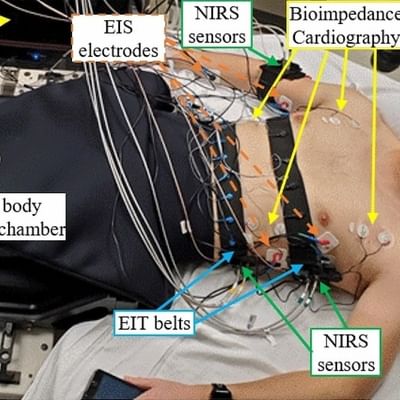
Necrotizing soft tissue infections (NSTIs) are aggressive infections that can progress rapidly from mild symptoms to sepsis, multi-organ failure, and death. NSTI cases present with non-specific clinical, imaging, and laboratory findings. Standard-of-care techniques for NSTI diagnosis lack sensitivity and specificity. A distinguishing histological feature of NSTIs is prominent blood vessel thrombosis in affected tissues. Leveraging this pro-thrombotic effect, our study group has demonstrated in a first-in-human study (NCT04839302) that intravenous administration of indocyanine green (ICG) and immediate fluorescence imaging reveals prominent signal deficits in NSTI-positive tissues. Our team is now leading a prospective, observational, multicenter clinical study to study video-rate ICG fluorescence imaging of patients suspected of having NSTIs who present to eight tertiary, Level 1 medical centers across the United States. The overall goal is to investigate whether ICG fluorescence imaging may act as a rapid diagnostic tool for NSTI, improving outcomes for patients with these life-threatening infections.
-
Quantitative scatter imaging
Quantitative scatter imaging
Quantitative scatter imaging makes use of the fact that normal functioning cells and cancerous cells show differences both in the components within the cells and in the structural organization of the cells within the tissue. These physical distinctions in biological structure have been shown to scatter light differently. This study develops a new approach to imaging scatter-based contrast over a wide field of view in tissue using high frequency structured light. These scattering features may provide a method to diagnostically identify abnormal tissue without the need to administer targeted compounds. This approach has the potential to generate new diagnostic screenings and new approaches for surgical guidance.
-
Photodynamic therapy
Photodynamic therapy
Photodynamic therapy (PDT) is a newly emerging therapy for displastic tissues, such as cancer, age-related blindness, pre-malignant transformation or psoriasis. The therapy involves the administration of a photosensitizing agent, together with the application of moderate intensity light to active the molecules to produce local doses of singlet oxygen. Ongoing research topics include, developing improved dosimetry instrumentation and software, fluorescence tomography imaging to sense drug localization, and assaying unique tumor biology and treatment effects in experimental cancers.
-
Fluorescence-guided surgery
Fluorescence-guided surgery
Fluorescence-guided surgery is important for the resection of some types of cancerous tumors where the tumor and normal tissue are similar in appearance and texture, and patient prognosis depends heavily on the completeness of resection. By selectively tagging tumor tissue with fluorescent dyes, it becomes possible to visually discriminate between normal and tumor tissues and improve significantly the completeness of tumor resection.
Selected Publications
- JT Elliott, K Marra, LT Evans, SC Davis, KS Samkoe, J Feldwisch, KD Paulsen, DW Roberts, BW Pogue. Simultaneous in vivo fluorescent markers for perfusion, protoporphyrin metabolism and EGFR expression for optically guided identification of orthotopic glioma. Clinical Cancer Res 2016; 10.1158/1078-0432.CCR-16-1400.
- JT Elliott, AV Dsouza, SC Davis, JD Olson, KD Paulsen, DW Roberts, BW Pogue. Review of fluorescence guided surgery visualization and overlay techniques. Biomed Opt Expr 2015; 6(10):3765–3782.
- JT Elliott, KS Samkoe, SC Davis, JR Gunn, KD Paulsen, DW Roberts, BW Pogue. Image-derived arterial input function for quantitative fluorescence imaging of receptor-drug binding in vivo. J Biophotonics 2015; DOI: 10.1002/jbio.201500162).
- AV DSouza, JT Elliott, JR Gunn, RJ Barth, KS Samkoe, KM Tichauer, Pogue BW. Nodal lymph flow quantified with afferent vessel input function allows differentiation between normal and cancer-bearing nodes. Biomed Opt Expr 2015: 6(4):1304–1317.
- SC Kanick, DM McClatchy III, V Krishnaswamy, JT Elliott, KD Paulsen, BW Pogue. Sub-diffusive scattering parameter maps recovered using wide-field high-frequency structured light imaging. Biomed Opt Expr 2014; 5(10):3376–90.
- JT Elliott, KS Samkoe, JR Gunn, Errol E. Stewart, Timothy B. Gardner, KM Tichauer, T-Y Lee, P. Jack Hoopes, Stephen P. Pereira, T Hasan, BW Pogue. Perfusion CT estimates photosensitizer quantification and biodistribution in a rabbit orthotopic pancreas cancer model. Academic Radiology 2014; 22(5):572–579.
- JT Elliott, M Diop, LB Morrison, CD. d'Esterre, T-Y Lee, K St. Lawrence. Quantifying cerebral blood flow in an adult pig ischemia model by a depth-resolved dynamic contrast-enhanced optical method. Neuroimage 2014; 94:303–11 DOI10.1016/j.neuroimage.2014.03.023.
- V Krishnaswamy, JT Elliott, DM McClatchy III, BW Pogue, KD Paulsen. Structured light scatteroscopy. Journal of Biomedical Optics 2014; 19(7):070504.
- JT Elliott, KM Tichauer, KS Samkoe, JR Gunn, KJ Sexton, and BW Pogue. Direct characterization of tracer plasma curves by fluorescence imaging of exposed carotid artery to facilitate kinetic analysis. Molecular Imaging in Biology 2014; DOI 10.1007/s11307-013-0715-y.
- KM Tichauer, M Diop, JT Elliott, Kimberly S. Samkoe, Tayabba Hasan, K St. Lawrence, and BW Pogue. Accounting for pharmacokinetic differences in dual-tracer receptor density imaging. Physics in Medicine and Biology 2014; 59(10):2341–51.
Patents
- System and method to detect the presence and progression of diseases characterized by systemic changes in the state of the vasculature | 12569149
- Method and apparatus to measure bone hemodynamics and discriminate healthy from diseased bone, and open reduction internal fixation implant with integrated optical sensors | 12257066