"Current methods aren't very specific," says Dartmouth engineering professor and lead author Jonathan Elliott, "The dyes often either show up in healthy tissue, or don't show up at the edges of tumors where they are most difficult to delineate. The results of this research show that ABY-029 can provide additional contrast and may greatly improve brain cancer resection rates."
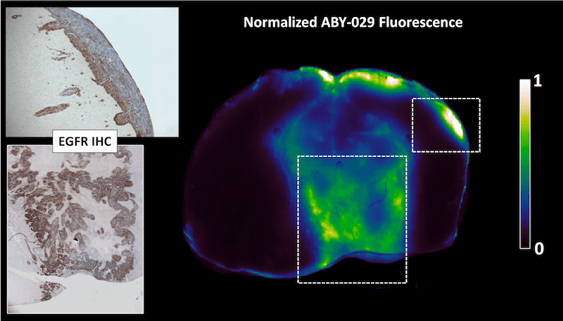
A color map representation of the ABY-029 fluorescence signal in an epidermal growth factor receptor (EGFR) tumor. Strong spatial agreement between ABY-029 fluorescence and EGFR-stained IHC images were observed in the tumor core and at distant regions of tumor invasion.
Fluorescence guided surgery is a technique that uses fluorescent dyes to highlight cancer during surgery, allowing the surgeon to home in on malignant tissue and remove it. Gliomas — any tumor that arises from the supportive (“gluey”) tissue of the brain — are notoriously difficult to remove completely, resulting in cancer recurrence and lower survival rates. ABY-029 targets epidermal growth factor receptor (EGFR), a molecule that is expressed on most glioma cells but not in normal brain tissue.
"The really exciting thing," continues Elliott, "is that the study brings us one step closer to providing patients and surgeons with personalized fluorescent dyes — being able to select the particular dye that will stick to their particular tumor — to achieve the most complete cancer removal for each patient."