- Undergraduate
Bachelor's Degrees
Bachelor of ArtsBachelor of EngineeringDual-Degree ProgramUndergraduate AdmissionsUndergraduate Experience
Quick Links
- Graduate
Graduate Experience
Quick Links
- Research
Research by Program Area
Quick Links
All Thayer News
Lab Report: Surgical Camera Pinpoints Tumors
Jul 01, 2022 | by Julie Bonette | Dartmouth Engineer
A new surgical camera developed by Dartmouth engineers and researchers at the Swiss Federal Institute of Technology Lausanne can image and measure the shape and location of deep-seated tumors—a great advancement on current operating room techniques.
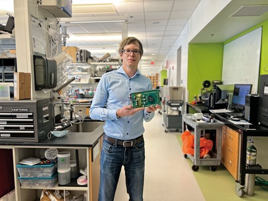
3-D SENSOR: Professor Petr Brůža holds a prototype of the fluorescence LiDAR camera. (Courtesy Photo)
Surgeons initially use computer tomography (CT) or magnetic resonance imaging (MRI) scans to gauge tumor locations and shapes, but during surgery typically rely on vision, touch, and training to remove tumors and leave healthy tissue intact. “We can use pulses of light to range the distance and position of cancerous cells in tissue, similar to how radar detects the position of planes in the air,” says Professor Petr Brůža.
The concept of using a depth-sensing camera—leveraging 3-D imaging sensor and pulses of light to localize tumors—came to Brůža in 2020. He assembled a team of biomedical engineers, including Professor Kimberley Samkoe, one of the principal investigators of Dartmouth’s fluorescence-guided surgery project, and Professor Edoardo Charbon in Switzerland, who provided an ultra-fast sensor for experimentation.
The team’s research—“Single-photon avalanche diode imaging sensor for subsurface fluorescence LiDAR”—was featured on the cover of Optica last winter. Now, Dartmouth engineers are testing a novel fluorescing drug designed to bind to cancer and highlight affected areas when irradiated by red light. The camera is being integrated into a compact surgical microscope to assist surgeons in navigating through sensitive tissue to ensure no tumor is left behind.
This article appeared in the Spring 2022 issue of the Dartmouth Engineer magazine.
For contacts and other media information visit our Media Resources page.