- Undergraduate
Bachelor's Degrees
Bachelor of ArtsBachelor of EngineeringDual-Degree ProgramUndergraduate AdmissionsUndergraduate Experience
Quick Links
- Graduate
Graduate Experience
Quick Links
- Research
Research by Program Area
Quick Links
All Thayer News
Dartmouth Engineers Develop Surgical Camera to Pinpoint Deep-Seated Tumors
Nov 12, 2021 | by Julie Bonette
A new surgical camera co-developed by Dartmouth engineers and researchers at the École polytechnique fédérale de Lausanne (EPFL) in Switzerland can image and measure the shape and location of deep-seated tumors.
Surgeons typically gauge tumor locations and shapes prior to surgery from computer tomography (CT) or magnetic resonance imaging (MRI) scans. But in the operating room, they rely heavily on vision, touch, and training to remove tumors and leave healthy tissue intact — an approach that's highly subjective to each surgeon.
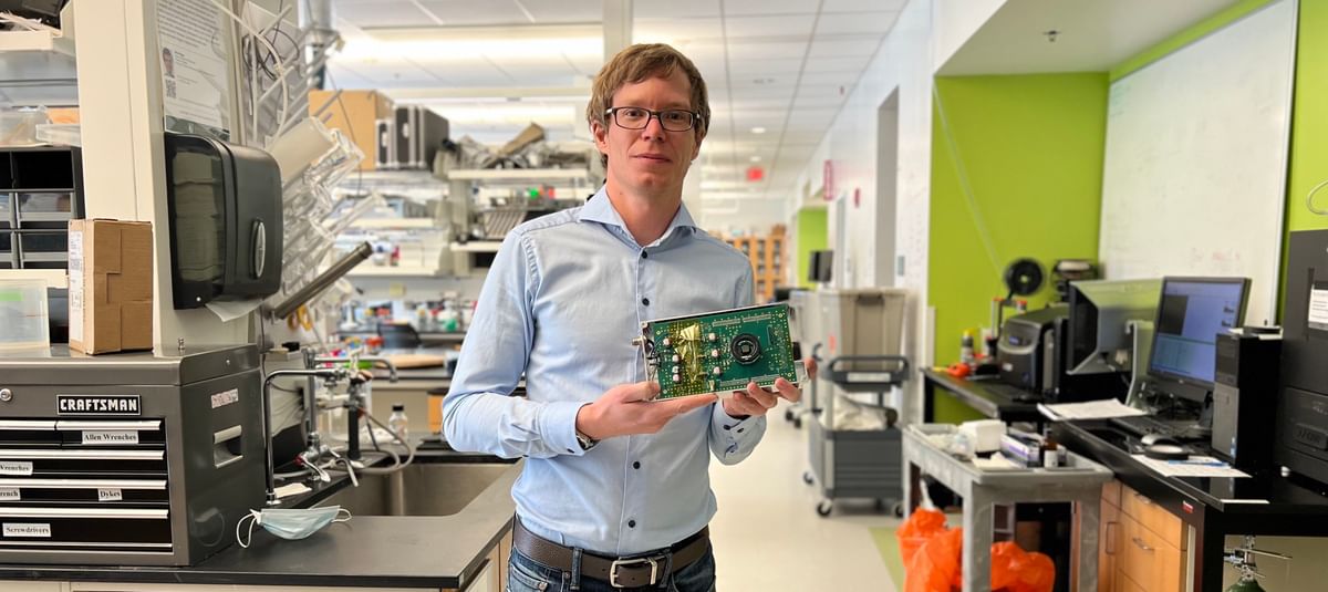
Dartmouth Engineering Assistant Professor Petr Brůža holds a prototype of the fluorescence LiDAR camera. (Photo by Rongxiao Zhang)
"We can use pulses of light to range the distance and position of cancerous cells in tissue, similar to how radar detects the position of planes in the air."
Professor Petr Brůža
Sub-surface LiDAR of a fluorescent labeled tumor with a large format single-photon avalanche diode array.
The idea for the depth-sensing camera came to Dartmouth Engineering Assistant Professor Petr Brůža a year ago, when he presented the concept of utilizing a 3D imaging sensor and pulses of light to localize tumors. For a long time, this posed a significant challenge as it only takes light less than a billionth of a second to travel back and forth in tissue.
In the paper, "Single-photon avalanche diode imaging sensor for subsurface fluorescence LiDAR," featured on the cover of Optica, Brůža proved this idea using the newest generation of imaging sensors. To do so, he assembled a team of biomedical engineers, including Professor Kimberley Samkoe, one of the principal investigators of Dartmouth's fluorescence-guided surgery project, as well as Edoardo Charbon, an EPFL professor, and his Advanced Quantum Architecture Laboratory team, which provided their ultra-fast sensor for experimentation.
Now, Dartmouth engineers are clinically testing a novel fluorescing drug designed to bind to cancer and highlight affected areas when irradiated by red light. The fluorescence LiDAR camera works alongside the drug by projecting red light pulses while simultaneously taking time-resolved pictures of the surface. When the red photons reach a deep tumor, it takes them longer to return to the surface. That time difference gives researchers the information they need to reconstruct the location and shape of the tumor.
The camera is currently being integrated into a compact surgical microscope, which will assist surgeons in navigating through sensitive tissue and confirming that no tumor is left behind after surgery. The researchers leveraged an ongoing first-in-human clinical trial of ABY-029, a targeted fluorescent peptide, to image oral head and neck cancer in an excised surgical specimen.
"This work would not be possible without the community of tremendously supportive doctors and clever engineers at Dartmouth and EPFL, as well as Prouty Pilot funding from Norris Cotton Cancer Center," said Brůža.
For contacts and other media information visit our Media Resources page.