- Undergraduate
Bachelor's Degrees
Bachelor of ArtsBachelor of EngineeringDual-Degree ProgramUndergraduate AdmissionsUndergraduate Experience
- Graduate
Graduate Experience
- Research
- Entrepreneurship
- Community
- About
-
Search

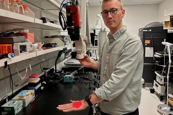
Pétusseau Th'23 demonstrates a new imaging technology, called "Pressure-Enhanced Sensing of Tissue Oxygenation," (PRESTO) that can help surgeons distinguish cancer from healthy tissue in real time. (Photo by Catha Mayor)
Overview
Arthur Pétusseau Th'23 earned dual master's degrees in engineering and science from the University of Strasbourg, France, in 2018 before completing his PhD in biomedical engineering at Dartmouth. His research focuses on developing innovative time-gated optical imaging technologies to enhance cancer diagnostics and treatment. Professor Pétusseau specializes in creating advanced imaging systems and minimally invasive diagnostic tools, prioritizing novel methodologies over commercial solutions. With expertise in hardware development and hands-on experience in living systems biology and animal surgery, he works to translate cutting-edge lab-born technologies into clinical applications.
Research Interests
Image-guided surgery; fluorescence imaging; molecular imaging; oximetry; radiotherapy; ultra-high dose rate radiotherapy; cancer research
Education
- MS, Engineering Sciences, Strasbourg 2018
- MEng, Engineering Sciences, Strasbourg 2018
- PhD, Engineering Sciences, Dartmouth 2023
Awards
- John W. Strohbehn, PhD, Memorial Prize
- Stu Trembly Award, Dartmouth Innovations Accelerator for Cancer
- 2nd place, Collegiate Inventors Competition
Startups
Hypoxia Surgical LLC
Co-Founder and CSO
Co-Founder and CSO
Research Projects
-
Exploring the role of oxygen in ultra high dose rate radiotherapy
Exploring the role of oxygen in ultra high dose rate radiotherapy
Ultra-high dose rate (UHDR) radiotherapy (RT) is an emerging cancer treatment that delivers high-energy beams (>40 Gy/s) to target tumors. Unlike conventional radiotherapy, which can damage healthy tissue, UHDR RT has been shown to spare normal tissue while maintaining tumor-killing effectiveness—a phenomenon known as the FLASH effect. The underlying mechanisms of this effect remain unclear. Our research focuses on investigating the role of oxygen in the FLASH effect using optical methods to measure tissue oxygen levels in real time during treatment.
Selected Publications
- Pétusseau AF, Ochoa M, Reed M, Doyley MM, Hasan T, Bruza P, Pogue BW (2024). Pressure-enhanced sensing of tissue oxygenation via endogenous porphyrin: Implications for dynamic visualization of cancer in surgery. Proceedings of the National Academy of Sciences 121(34): e2405628121.
- Pétusseau AF, Clark M, Bruza P, Gladstone D, Pogue BW (2024). Intracellular oxygen transient quantification in vivo during ultra-high dose rate FLASH radiation therapy. International Journal of Radiation Oncology, Biology, Physics. Advance online publication. https://doi.org/10.1016/j.ijrobp.2024.04.068
- Pétusseau A, Bruza P, Pogue B (2022). Protoporphyrin IX delayed fluorescence imaging: A modality for hypoxia-based surgical guidance. Journal of Biomedical Optics 27(10): 106005. https://doi.org/10.1117/1.JBO.27.10.106005
- Pétusseau AF, Streeter SS, Ulku A, Feng Y, Samkoe KS, Bruschini C, Charbon E, Pogue BW, Bruza P (2024). Subsurface fluorescence time-of-flight imaging using a large-format single-photon avalanche diode sensor for tumor depth assessment. Journal of Biomedical Optics 29(1): 016004. https://doi.org/10.1117/1.JBO.29.1.016004
- Scholz M, Pétusseau AF, Gunn JR, Chapman MS, Pogue BW (2020). Imaging of hypoxia, oxygen consumption, and recovery in vivo during ALA-photodynamic therapy using delayed fluorescence of protoporphyrin IX. Photodiagnosis and Photodynamic Therapy 30. https://doi.org/[Insert DOI]
- Brůža P, Pétusseau A, Tisa S, Jermyn M, Jarvis LA, Gladstone DJ, Pogue BW (2019). Imaging Cherenkov photon emissions in radiotherapy with a Geiger-mode gated quanta image sensor. Optics Letters 44(18): 4546–4549. https://doi.org/[Insert DOI]


