- Undergraduate
Bachelor's Degrees
Bachelor of ArtsBachelor of EngineeringDual-Degree ProgramUndergraduate AdmissionsUndergraduate Experience
Quick Links
- Graduate
Graduate Experience
Quick Links
- Research
Research by Program Area
Quick Links
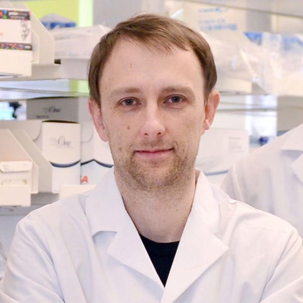
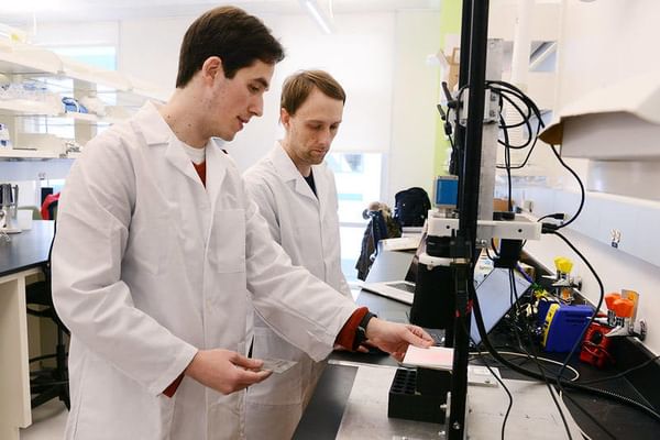
Professor Davis with PhD candidate Ethan Larochelle at the Williamson Translational Research Building. (Photo by Kathryn LoConte Lapierre)
Research Interests
Engineering in medicine; cancer diagnostics and treatment; biomedical optics; multi-modal medical imaging; molecular imaging; fluorescence molecular tomography; fluorescence guided surgery; photodynamic therapy
Education
BA, Physics, Middlebury College 1997
MSc, Mechanical Engineering, University of Virginia 2000
PhD, Engineering Sciences, Dartmouth College 2008
Awards
- Department of Defense Breast Cancer Research Program Post-doctoral Traineeship Award
- Department of Defense Breast Cancer Research Program Pre-doctoral Traineeship Award
- Thomas Kelvin Burnap Fellowship, Dartmouth College
- Dean’s Scholarship, University of Virginia
- Chairman’s Scholarship, University of Virginia
- Phi Beta Kappa
Professional Activities
- Dartmouth College Irradiation Safety Committee
- World Molecular Imaging Society (WMIS)
- Society of Photo-optical Instrumentation Engineers (SPIE)
- Optical Society of America (OSA)
- American Society of Photobiology (ASP)
Startups
DoseOptics LLC
Co-founder and Managing Member
Co-founder and Managing Member
Research Projects
-
Cerenkov imaging in radiation therapy
Cerenkov imaging in radiation therapy
Radiation therapy is used to treat cancer tumors by killing the tissue with high ionizing radiation doses. Until recently it has not been possible to image the radiation dose delivered to tissue, but through Cherenkov light imaging, this delivered dose can be mapped with high resolution cameras. The research group focuses on quantification of the imaging, and developing tools which allow radiation therapy to be delivered in a safer and more validated manner.
-
Photodynamic therapy
Photodynamic therapy
Photodynamic therapy (PDT) is a newly emerging therapy for displastic tissues, such as cancer, age-related blindness, pre-malignant transformation or psoriasis. The therapy involves the administration of a photosensitizing agent, together with the application of moderate intensity light to active the molecules to produce local doses of singlet oxygen. Ongoing research topics include, developing improved dosimetry instrumentation and software, fluorescence tomography imaging to sense drug localization, and assaying unique tumor biology and treatment effects in experimental cancers.
-
Fluorescence-guided surgery
Fluorescence-guided surgery
Fluorescence-guided surgery is important for the resection of some types of cancerous tumors where the tumor and normal tissue are similar in appearance and texture, and patient prognosis depends heavily on the completeness of resection. By selectively tagging tumor tissue with fluorescent dyes, it becomes possible to visually discriminate between normal and tumor tissues and improve significantly the completeness of tumor resection.
-
Optical molecular imaging
Optical molecular imaging
Optical molecular imaging is being used to provide molecular guidance in cancer surgery. Fluorescent contrast agents are in pre-clinical and clinical studies to image cancer tumors in vivo, with a dual focus, first on getting more accurate information out of the tissue, and secondly to provide better information about the specificity of the molecules as markers. Systems and algorithms for diffuse fluorescence imaging of tissue are studied, both as a stand-alone system, and as coupled to magnetic resonance imaging and computed tomography imaging. Tracer kinetic modeling is also being developed to allow quantitative imaging of molecular binding in vivo.
-
Near-infrared imaging
Near-infrared imaging
Near-infrared imaging (NIR) provides a way to quantify blood and water concentrations in tissue, as well as structural and functional parameters. Since normal tissue, benign tumors, and malignant tumors each carry different concentrations of both hemoglobin and water, and have different levels of oxygen demand and ultrastructural scattering, NIR spectroscopy can be combined into standard imaging systems as an effective method of to provide additional information for breast cancer detection and diagnosis. Work is ongoing to improve techniques for better image reconstruction, display and integration with magnetic resonance imaging (MRI) and computed tomography (CT) imaging.
See also Center for Imaging Medicine
Selected Publications
- Kanick SC*, Davis SC*, Zhao Y, Hasan T, Maytin EV, Pogue BW, Chapman MS, “Dual channel red/blue fluorescence dosimetry with broadband reflectance spectroscopic correction measures protoporphyrin IX production levels during PDT of actinic keratosis,” Journal of Biomedical Optics 19(7): 75002 (2014). *denotes equal contributions.
- Jermyn M, Davis SC, Dehghani H, Huggett M, Hasan T, Pereira SP, Pogue BW, “CT contrast predicts pancreatic cancer treatment response to verteporfin-based photodynamic therapy,” Physics in Medicine and Biology 59(8): 1911-22 (2014).
- Davis SC, Gibbs SL, Gunn JR, Pogue BW, “Topical dual-stain difference imaging for rapid intra-operative tumor identification in fresh specimens,” Optics Letters 38(23): 5184-5187 (2013).
- Davis SC, Samkoe KS, Tichauer KM, Sexton KJ, Gunn JR, Deharvengt SJ, Hasan T, Pogue BW, “Dynamic dual-tracer MRI-guided fluorescence tomography to quantify receptor density in vivo,” Proceedings of the National Academy of Sciences 110(22): 9025-9030 (2013). PMC3670304
- Zhang R*, Davis SC*, Demers JL, Glaser AK, Vinogradov S, Gladstone DJ, Pogue BW, “Intensity and Lifetime Reconstruction of Oxygenation-Sensitive Phosphorescence Excited by Čerenkov Radiation during EBRT,” (* denotes equal contributions) Journal of Biomedical Optics Letters 18(5):050503 (2013). PMC3643897
- Axelsson J, Davis SC, Gladstone DJ, Pogue BW, “Cherenkov emission induced by external beam radiation stimulates molecular fluorescence,” Medical Physics Letters 38(7):4127-4132 (2011). PMC3139507
- Leblond F, Davis SC, Valdes PA, Pogue BW, “Preclinical whole-body fluorescence molecular imaging: Review of instruments, methods and applications,” Journal of Photochemistry and Photobiology B: Biology 98(1):77-94 (2010). PMC3678966
- Davis SC, Pogue BW, Springett R, Leussler C, Mazurkewitz P, Tuttle SB, Gibbs-Strauss SL, Jiang S, Dehghani H, , Paulsen KD, “Magnetic resonance-coupled fluorescence tomography scanner for molecular imaging of tissue,” Review of Scientific Instruments, 79-064302-1-10 (2008). PMC2678791
- Dehghani H, Davis SC, Jiang SD, Pogue BW, Paulsen KD, Patterson MS, “Spectrally resolved bioluminescence optical tomography,” Optics Letters, 31(3):365–367 (2006).
- Pogue BW, Davis SC, Song XM, Brooksby BA, Dehghani H, Paulsen KD, “Image analysis methods for diffuse optical tomography,” J. Biomed Optics, 99(3):033001:1-12 (2006).
Patents
- Methods for quantitative and enhanced-contrast molecular medical imaging using cross-modality correction for differing tracer kinetics | 12144667
News


In the News
The New York Times
Biden Awards $150 Million in Research Grants as Part of Cancer 'Moonshot'
Aug 14, 2024
Biden Awards $150 Million in Research Grants as Part of Cancer 'Moonshot'
Aug 14, 2024