- Undergraduate
Bachelor's Degrees
Bachelor of ArtsBachelor of EngineeringPartner School Dual-DegreeUndergraduate AdmissionsUndergraduate Experience
Quick Links
- Graduate
Doctoral Degrees
Doctor of PhilosophyPhD Innovation ProgramDoctor of Medicine-PhDGraduate AdmissionsGraduate Experience
Quick Links
- Research
Research by Program Area
Quick Links
All Thayer News
New Imaging System Makes Back Surgery Safer, Faster and Less Expensive
Apr 25, 2018
Dartmouth engineers and physicians developed an innovative intraoperative stereovision system.
Researchers at Dartmouth have found a way to make back surgery safer, faster and more cost effective.
MRIs and CT scans help surgeons identify spine problems, like compressed vertebrae or herniated disks, but finding a clear path to those problem areas is not always as straightforward. Tissue and bone not only stand in the way, they can also move during spinal surgery, rendering a CT scan taken prior to surgery much less accurate.
To solve this problem, Dartmouth professors from Thayer School of Engineering and Geisel School of Medicine developed a 3-dimensional, real-time optical tracking system to guide back surgeons as they operate, like a Google Maps for the body, according to findings published in the journal Operative Neurosurgery.
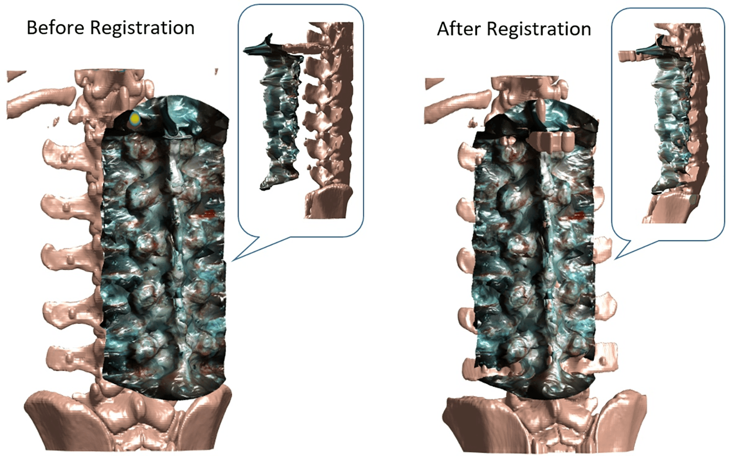
Registration of CT (computed tomography) and iSV (intraoperative stereovision). CT is transformed to match its surface to iSAV, represented in tracking system coordinates.
Using a complex software algorithm and two cameras attached to a surgical microscope, the system produces real-time 3-dimensional digitized images on a monitor, according to the study. This type of tracked, calibrated stereoscopic camera system has been extensively used in brain surgery but until now has been unexplored for use in spinal surgery.
The surgeon can use this new intraoperative stereovision system (iSV) without any additional radiation or labor-intensive marking of key areas on the patient’s spine, to match up or co-register with the pre-operative CT scan, as some surgeons do today. This new mapping provides more accurate renderings of where spinal implants or other surgical tools and devices need to go during the procedure, and is expected to save up to 30 minutes, according to one of the study’s authors, Keith Paulsen, The Robert A. Pritzker Professor of Biomedical Engineering at Thayer School of Engineering at Dartmouth.
Paulsen and the multidisciplinary team at Dartmouth’s Center for Surgical Innovation tested the new iSV system for accuracy and efficiency while operating on pig spines. Since completing this study, the team has taken its complex system one step further by converting it into a handheld “wand” that the surgeon can pass over the surgical area.
"By rendering images real-time, with a simple handheld tool, we believe we can make surgeries safer and less costly in the future," said Paulsen.
Next up is fine-tuning the system and testing in humans. The National Institutes of Health has provided the Dartmouth team with another round of funding to continue testing. It could be several years before the system becomes widely available for human spinal surgeries.
Media Contact: Callaway Zuccarello; 314.862.4300
The findings are shared in the journal Operative Neurosurgery in a paper co-authored by Linton Evans, MD, Jonathan D. Olson, BS, Yunliang Cai, PhD, Xiaoyao Fan, PhD, Keith D Paulsen, PhD, David W. Roberts, MD, Songbai Ji, ScD, and S. Scott Lollis, MD.
Research reported in this publication was supported by the National Institute of Biomedical Imaging and Bioengineering of the National Institutes of Health under Award Numbers R01EB025747 and T32EB02196. The content is solely the responsibility of the authors and does not necessarily represent the official views of the National Institutes of Health.
Operative Neurosurgery, opy023, https://doi.org/10.1093/ons/opy023
For contacts and other media information visit our Media Resources page.