- Undergraduate
Bachelor's Degrees
Bachelor of ArtsBachelor of EngineeringPartner School Dual-DegreeUndergraduate AdmissionsUndergraduate Experience
Quick Links
- Graduate
Doctoral Degrees
Doctor of PhilosophyPhD Innovation ProgramDoctor of Medicine-PhDGraduate AdmissionsGraduate Experience
Quick Links
- Research
Research by Program Area
Quick Links

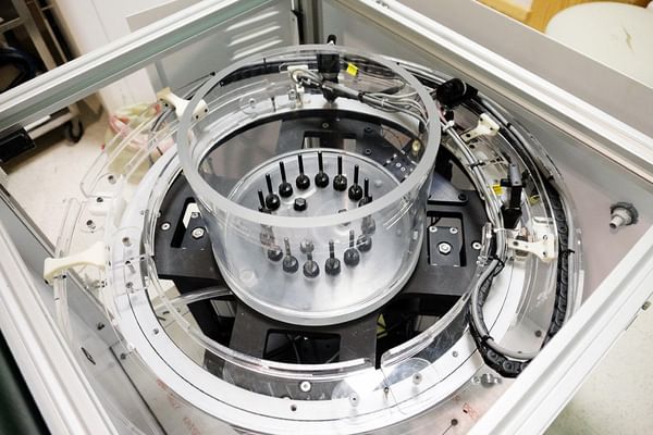
Professor Meaney is part of a team developing a multi-modal breast imaging platform that uses both microwave and magnetic resonance imaging, a first in the field. (Photo by Shireen Geimer)
Research Interests
Microwave imaging ultrasound computed tomography for biomedical applications; microwave antenna design; thermal modeling and system design for focused ultrasound surgery applications
Education
- AB, Electrical Engineering and Computer Science, Brown University 1982
- MS, Electrical Engineering, University of Massachusetts 1985
- PhD, Engineering Sciences, Dartmouth College 1995
Professional Activities
- Associate Editor, IEEE Transactions on Biomedical Engineering
- Member, B-CRATOS Advisory Board
Research Projects
-
Microwave imaging spectroscopy
Microwave imaging spectroscopy
Microwave imaging spectroscopy (also commonly referred to as microwave computed tomography) presents the challenge of measuring the signal data necessary to produce meaningful images. Development of site-specific antenna arrays along with improved electronic signal detection technology is rapidly making this measurement feasible for breast cancer detection. A second-generation tomographic breast imaging system has been completed and the data are being used to recover permittivity and conductivity maps of the breast for evaluation by a clinician.
-
Therapy monitoring
Therapy monitoring
Therapy monitoring is an important emerging application of imaging modalities. These and other current research topics include:
- near-infrared imaging of brain tissue;
- near-infrared spectroscopy for diagnosing peripheral vascular disease;
- electrical impedance spectroscopy for radiation therapy monitoring;
- magnetic resonance elastography for detecting brain or prostate lesions; to follow the progression of diabetic damage in the foot; and to answer basic questions of wave propagation in tissue;
- microwave imaging spectroscopy for hyperthermia therapy monitoring, brain imaging, and detection of early-stage osteoporosis;
- electrical impedance tomography for monitoring traumatic brain injury progression and therapy.
-
Non-linear image reconstruction techniques
Non-linear image reconstruction techniques
Non-linear image reconstruction techniques is at the core of the medical imaging projects. Excitation-induced measurements from each instrument are compared with calculations from corresponding numerical models to compute updated property images of the biological target. As the images are progressively updated (or refined) in a non-linear iterative process, important features and functional information related to the objects physiological status—tumor, benign tissue, etc.—become more apparent. The computational core of the breast imaging project works synergistically with all four groups to improve our fundamental understanding of these mathematical systems to improve overall image quality and resolution. These processes have been developed for both 2D and 3D geometries in each modality and are being expanded to exploit emerging parallel computing capabilities.
-
Microwave electronics
Microwave electronics
Microwave electronics capable of fast data acquisition (approaching real-time) are being developed for brain imaging applications. Also, site-specific antenna arrays for microwave imaging are in development for heel imaging (screening for osteoporosis) and for brain imaging applications.
-
Dielectric properties of tissue
Dielectric properties of tissue
Dielectric properties of tissue—measured through advanced microwave imaging techniques—convey functional information useful for making clinical diagnoses. The properties reflect tissue composition of fat, bone, water, proteins, etc., and often have unique spectral characteristics. The relative proportions and dynamic aspects of these constituents can have important implications for breast cancer imaging, osteoporosis detection, brain imaging, and heat therapy monitoring.
Patents
- Imaging by magnetic resonance adsorption, elastography and tomography | 8,207,733
- Microwave imaging system and processes, and associated software products | 7,825,667
- Non-invasive microwave analysis methods | 7,755,010
- Magnetic resonance microwave absorption and tomography | 7,439,736
- Non-invasive microwave analysis systems | 7,319,212
- Non-invasive microwave analysis systems | 7,164,105
- Fixed array imaging, high contrast object imaging techniques and breast cancer imaging | 6,448,788
- Two-dimensional microwave imaging apparatus and methods | 5,841,288
Courses
- ENGS 7: First Year Seminars in Engineering
Books

Alternative Breast Imaging: Four Model-Based Approaches
Springer, 2004
Videos
B-CRATOS Project Interview
News


